A persistently small, constricted pupil is primarily mediated by parasympathetic nervous system activity. This condition, referred to as pupillary miosis, may result from a variety of causes, including opioid or narcotic use, antihypertensive medications, Horner’s syndrome, traumatic brain injury, anterior uveitis, and exposure to certain pesticides.
Pupillary constriction, or miosis, results from disruption or irritation of the autonomic innervation of the pupils. It is defined by a pupil diameter of 2.5 mm or less, corresponding to less than 21% of the iris diameter. Miosis is generally classified into two forms: paralytic miosis, caused by impairment of the pupillary dilator muscle due to disruption of the sympathetic pathways, and spastic miosis, resulting from excessive contraction of the pupillary sphincter secondary to irritation of the parasympathetic pathways.
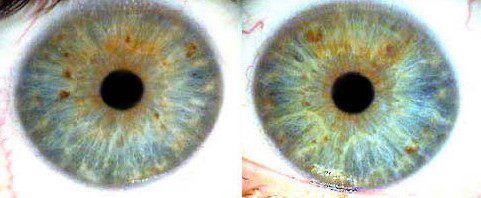
Physiological Miosis
Physiological miosis may be influenced by several intrinsic and functional factors, including:
- Constitutional characteristics, such as increased iris pigmentation, parasympathicotonia, and the relative balance between cholinergic and adrenergic systems.
- Age-related changes, reflecting a decline in adaptive and protective mechanisms, including iris atrophy.
- Vagotonic influences, characterized by transient parasympathicotonia associated with mental or physical fatigue, hyperventilation, sleep, or postprandial states.
- Psycho-emotional factors, including heightened stress or aggressive states.
- Refractive status, with pupils typically narrower in hypermetropic individuals than in emmetropes, and narrower in emmetropes than in myopes.
Pathological Miosis
Pathological miosis may arise from conditions affecting central autonomic regulatory centers, including:
- Exogenous intoxications, such as exposure to pharmacological agents (e.g., carbocholine, pilocarpine, eserine, pyrophosphate, morphine), alcohol, or carbon monoxide.
- Endogenous intoxications associated with comatose states, including uremic, diabetic, alimentary, and dystrophic coma.
- Astheno-depressive psychotic states.
- Acute cerebrovascular disorders.
- Hypofunction of the autonomic nervous system, often accompanied by dysfunction in other organ systems, particularly the gastrointestinal tract.
Diagnostic Significance
Unilateral miosis is of particular diagnostic importance in the evaluation of central nervous system injury, especially involving the vertebrobasilar circulation.
Primary and secondary syndromes commonly associated with such lesions include Bernard–Horner syndrome, Dejerine–Klumpke syndrome, Pancoast syndrome, Babinski–Nageotte syndrome, and the alternating bulbar Wallenberg–Zakharchenko syndrome.