PupilMetrics Research is a clinical pupillometry platform designed for neurological assessment and TBI recovery monitoring. It turns the human pupil – the only visible part of the central nervous system – into a quantitative, repeatable, and objective diagnostic tool. By measuring the pupillary light reflex (PLR) with high precision, PupilMetrics Neuro helps neurologists, concussion clinics, sports medicine physicians, and researchers establish baselines, track recovery progress, detect subtle autonomic changes, monitor medication effects, and identify bilateral asymmetries (anisocoria).
Built on the core clinical premise that if the brain is healing, the pupil will show it, the system delivers research-grade measurements in a practical, non-invasive clinical workflow. It requires no blood draws, radiation, sedation, or active patient cooperation beyond opening the eyes. All processing occurs locally with no cloud connectivity required.
Main Features:
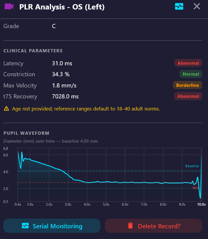
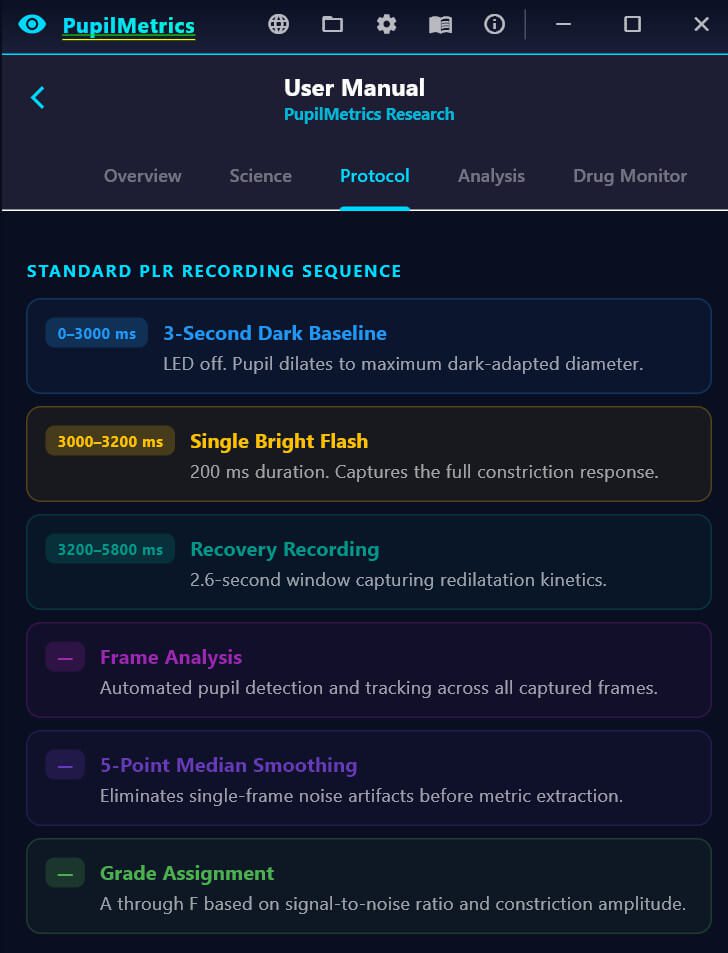
Structured PLR Recording & Analysis: Uses a precise protocol with a 3-second dark-adapted baseline, single bright flash, and automated pupil tracking. Delivers key metrics including baseline diameter, constriction amplitude, percent constriction, latency, velocity, redilatation speed, and an overall PLR grade (A–F).
3-Trial Habituation Protocol: Runs three consecutive PLR trials to calculate a habituation index, revealing cortical modulation and frontal-midbrain circuit integrity. Includes waveform overlay for visual comparison.
Bilateral Comparison & Anisocoria Detection: Automatically measures differences between right and left eyes and flags clinically significant asymmetry with severity grading.
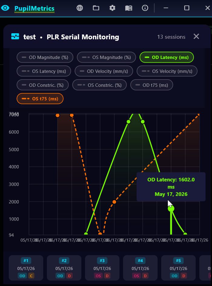
Serial Trend Monitoring: Enables clinicians to load past scans and track recovery trajectories over time (diameter normalization, PLR amplitude improvement, anisocoria resolution, and habituation normalization). Results are timestamped and exportable to PDF.
Dark-Adapted Baseline & Real Timestamps: Uses a 3-second complete dark period for maximum dynamic range and embeds actual wall-clock timestamps in every frame to ensure accurate, research-grade timing regardless of frame-rate variability.
Targeted Clinical Applications: Optimized for concussion/sports medicine clinics, neurology/neurotrauma, and clinical research. Supports sideline baseline testing, return-to-play decisions, ICU monitoring, pharmacodynamic studies, and longitudinal documentation.
Modern, Secure Technology Stack: Built with Flutter (cross-platform UI), Dino-Lite digital iriscope, ONNX Runtime for hybrid pupil detection, and local SQLite storage. Fully offline operation with complete patient data privacy.
Download and Install PupilMetrics Windows
PupilMetrics software is certified under the Microsoft Developer Program to ensure trust and safety!

If you run into any other issues during install or have questions about the tool, feel free to reply, we’re here to help! Thanks for using PupilMetrics in your research work!