The apparent color of the pupil is determined by the optical properties of the lens and the anterior chamber of the eye. Alterations in these structures may reflect dysfunction within broader physiological systems, including the endocrine and vascular systems. Any deviation of the pupil from its normal black appearance is regarded as an indicator of pathological change.
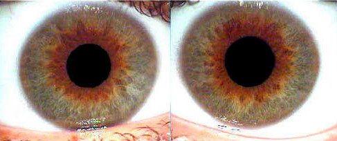
The most common cause of loss of the pupil’s normal black appearance is cataract formation. In these cases, the pupil may appear muddy gray to bright white and can present with various morphological patterns, including diffuse whitening, localized subcapsular opacities, central punctate lesions, radial striations, or sector-shaped bands. Under oblique illumination, these changes may be accompanied by alterations in iris coloration.
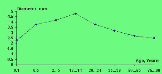
This curve illustrates age-related changes in pupil diameter.
Under physiological conditions, pupil diameter in darkness typically ranges from 2.7 to 4.8 mm (approximately 22.5%–40% of the iris diameter), with a mean value of 3.5 mm (about 29% of the iris diameter). In newborns, pupils are physiologically smaller, with an average diameter of 2.32 ± 0.05 mm, corresponding to 19%–20% of the iris diameter. Beginning in the second month of life, pupil size increases progressively, enlarging by approximately 64% by six months of age and by an additional 8.6% by the end of the first year. Growth continues until approximately 10–11 years of age.
From adolescence onward, a gradual age-related reduction in pupil diameter is observed, occurring in three distinct phases:
- Phase I (15–24 years): A marked decrease in pupil diameter of approximately 17%–19%.
- Phase II (25–50 years): A slower rate of constriction, with a reduction of approximately 6%–9% every five years.
- Phase III (50–80 years): Pupils remain relatively small, averaging 2.63 ± 0.06 mm (approximately 22% of the iris diameter), with a further gradual decrease of about 3%–4% per decade.
Pupillary tone—reflecting the balance between tension and relaxation—is regulated by the central nervous system in coordination with the autonomic nervous system. Reduced pupillary tone, manifested as relative dilation, may indicate general physical fatigue, whereas pupillary constriction is commonly associated with increased physiological or psychological tension. Pupillary dynamics are influenced by both physical and emotional factors, including fear, pain, excitement, boredom, and fatigue.
An understanding of age-related variations in pupil size is essential for distinguishing physiological changes from pathological forms of miosis and mydriasis.