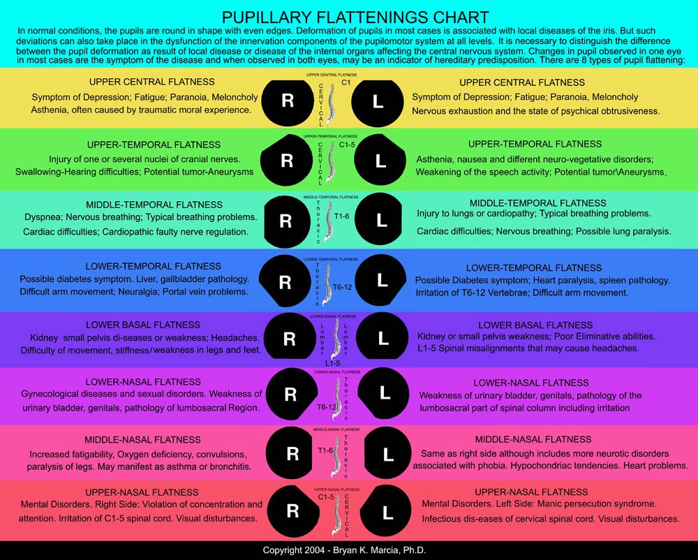
Under physiological conditions, pupils are round and have smooth, regular margins. Pupillary deformities are most commonly associated with localized iris pathology but may also result from disturbances in the innervation of the pupilomotor system at various levels. It is therefore essential to distinguish deformations caused by primary ocular disease from those arising secondary to systemic conditions affecting the central nervous system.
Unilateral alterations in pupil shape are generally indicative of an underlying pathological process, whereas bilateral changes may reflect a hereditary or constitutional predisposition. Multiple distinct types of pupillary deformation have been described. Deformations caused by partial loss or “melting” of the pupillary margin, rather than displacement of the pupillary stromal ring, are classified as false variants.
When assessed using slit-lamp biomicroscopy, pupillary flatness involving one-sixth or more of the pupillary circumference—corresponding to approximately two clock-face hours—is considered diagnostically significant. Advanced computer-based analysis enables substantially greater measurement precision, detecting pupillary contour variations as small as 0.1 clock-face hours, representing up to a fourfold increase in sensitivity compared with conventional examination methods.