The Pupils
The pupil is the central opening of the iris, shaped by the circular muscle and bordered by the pupillary margin, which regulates the amount of light entering the inner structures of the eye.
The condition and responses of the pupil reflect the functional state of various brain structures. Information about both afferent (somatic) and efferent (autonomic) components of cranial-cerebral innervation and the pupilomotor systems is combined, making the pupil a valuable and accessible indicator for assessing brainstem activity.
Pupil reflexes are crucial in diagnosing many neurological conditions, often as part of well-known syndromes such as Bernard-Horner, Adie’s, Argyll-Robertson, and Parinoud’s. Changes in pupil color, size, shape, position, and reaction are all clinically significant.
Pupil Deformation
Under normal conditions, pupils are round with even edges. Pupil deformation is often linked to local iris diseases, but it can also result from dysfunctions in the pupilomotor system’s innervation at any level. It is important to distinguish between deformation caused by local dystrophic processes and those that develop reflexively due to internal organ diseases or central nervous system conditions. The main types of pupil deformation include:
- Drawing (oval or elliptical forms)
- Local Flatness (sector deformations)
- Multiformity
When observed in one eye, these deformations often indicate pathology, while when seen in both eyes, they may suggest a hereditary predisposition.
Deformations caused not by a shift in the pupillary stroma but by the “melting” of parts of the pupillary margin are considered false variants.
Studies have shown that these changes may be related to symptoms of organs connected to reflexogenic zones in the iris adjacent to the deformed areas or in the same direction (e.g., ciliary area). Multiform deformations are commonly found in patients with brain tumors.
Pupillary flatness that affects at least one-sixth of the pupil’s circumference is significant for indicating active pathology.
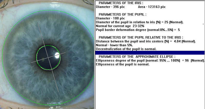
We use a comprehensive algorithmic data analysis to evaluate various pupil parameters, including miosis, mydriasis, anisocoria, and different forms of pupil deformation such as drawing, oval-elliptic shapes, local flatness, sector deformations, decentrations, and multiformitie.