Facebook Twitter VKontakte
Sign in
  • Home
  • Human Eye Project
    • The Pupil
    • Pupil Decentration-Multiformaties
    • Pupil Deformations
    • Pupil Color and Dimensions
    • Pupil Anisocoria
    • Pupil Miosis
    • Pupil Mydriasis
    • Pupil Reflexes
    • The Collarette
    • The Iris
  • PupilMetrics App
    • PupilMetrics Android
    • PupilMetrics Windows
    • PupilMetrics Mac OS
  • Documentation
  • Pricing
  • Learn More
    • CNRI Research
    • History
    • About
    • Eye Photo Tips & Tricks
    • IRB Status
    • Pupil Abstract Blog
    • Privacy Policy
    • Terms of Service
Sign in
Welcome!Log into your account
Forgot your password?
Password recovery
Recover your password
Search
Sign in
Welcome! Log into your account
Forgot your password? Get help
Password recovery
Recover your password
A password will be e-mailed to you.
Clinical Neuro-Optic Reseach Initiative
  • Home
  • Human Eye Project
    • The Pupil
    • Pupil Decentration-Multiformaties
    • Pupil Deformations
    • Pupil Color and Dimensions
    • Pupil Anisocoria
    • Pupil Miosis
    • Pupil Mydriasis
    • Pupil Reflexes
    • The Collarette
    • The Iris
  • PupilMetrics App
    • PupilMetrics Android
    • PupilMetrics Windows
    • PupilMetrics Mac OS
  • Documentation
  • Pricing
  • Learn More
    • CNRI Research
    • History
    • About
    • Eye Photo Tips & Tricks
    • IRB Status
    • Pupil Abstract Blog
    • Privacy Policy
    • Terms of Service

Getting Started

5
  • 1.1 System Requirements
  • 1.2 Installation
  • 1.3 Licensing
  • 1.4 First Launch
  • 1.5 Desktop Window & Keyboard Shortcuts

Capturing Eye Images

8
  • 2. Capturing Eye Images
  • 2.1 Camera Source Selection
  • 2.2 Quality-Gated Camera Mode – Android App
  • 2.3 Manual Camera Mode – Android
  • 2.4 USB / UVC Iriscope (Dino-Lite)
  • 2.5 PLR Video Mode – Android
  • 2.6 Import from Gallery
  • 2.7 Tips for a Good Capture

Reading the Analysis Results

10
  • 3. Reading the Analysis Results
  • 3.1 The Iris Zone Map
  • 3.2 PI Ratio (Pupil–Iris Ratio)
  • 3.3 Zone Findings — Flattenings (FLAT) and Protrusions (PROT)
  • 3.4 ANW Assessment (Collarette / Autonomic Nerve Wreath)
  • 3.5 Decentration (Pupil Position)
  • 3.6 Ellipseness (Pupil Shape)
  • 3.7 Anisocoria (Pupil Size Difference)
  • 3.8 Confidence Scores & Hybrid Fusion
  • 3.9 Scan History

Patient Management

4
  • 4. Patient Management & Exports
  • 4.1 PDF Report
  • 4.2 Plain-Text & JSON Export
  • 4.3 Sharing & Filing

Natural Medicine Therapy Panels

8
  • 5. Natural Medicine Therapy Panels
  • 5.1 Enabling the Therapy Modules
  • 5.2 How Zone Findings Drive the Therapy Panels
  • 5.3 Herbal Recommendations Panel
  • 5.4 Nutrition Recommendations Panel
  • 5.5 Chiropractic Correlations Panel
  • 5.6 TCM Correlations Panel
  • 5.7 Reading Therapy Panels Together

Constitutional Iridology

7
  • 6. Constitutional Iridology
  • 6.1 Background & Theoretical Basis
  • 6.2 The 34 Constitutional Types
  • 6.3 Selecting a Constitutional Type
  • 6.4 Constitutional Panel in Analysis Results
  • 6.5 Constitutional Section in the PDF Report
  • 6.6 Clinical Guidance & Limitations

Exporting PDF Reports

2
  • 7. Exporting PDF Reports
  • 7.1 Regenerating a PDF

Settings & Customization

5
  • 8. Settings & Customization
  • 8.1 Languages
  • 8.2 Zone Overlay & Observer Notes
  • 8.3 ML Comparison Panel
  • 8.4 About & Support

Clinical & Legal Disclaimers

2
  • 9. Clinical & Legal Disclaimers
  • 9.1 Data Privacy
View Categories
  • Home
  • PupilMetrics Documentation
  • Natural Medicine Therapy Panels
  • 5.5 Chiropractic Correlations Panel

5.5 Chiropractic Correlations Panel

1 min read

**Data source**

Spinal segment data is compiled directly into the app (no external asset file is loaded at runtime). The zone-to-segment mapping follows the **pupillary-border iridology convention**:

| Iris clock position | Spinal region |

|——————–|—————|

| 12 o’clock (upper-central) | Upper Cervical C1–C4 |

| 10–11 / 1–2 o’clock (upper) | Mid/Lower Cervical C4–C7 |

| 9 / 3 o’clock (middle) | Upper Thoracic T1–T6 |

| 7–8 / 4–5 o’clock (lower) | Lower Thoracic T7–T12 |

| 6 o’clock (lower-basal) | Lumbar / Sacral L1–S3 |

Both eyes map to the **same spinal column** — the spine is midline, so OD and OS findings at the same clock position point to the same vertebral level.

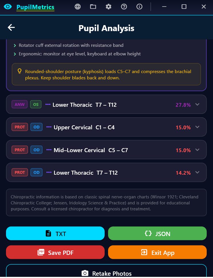
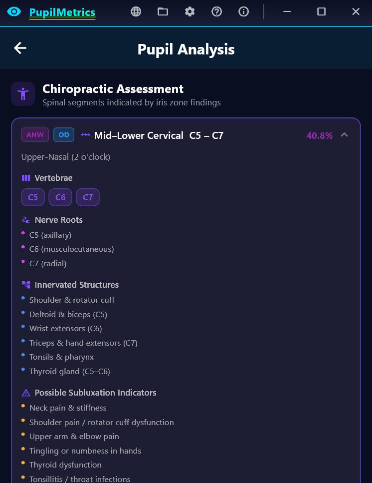
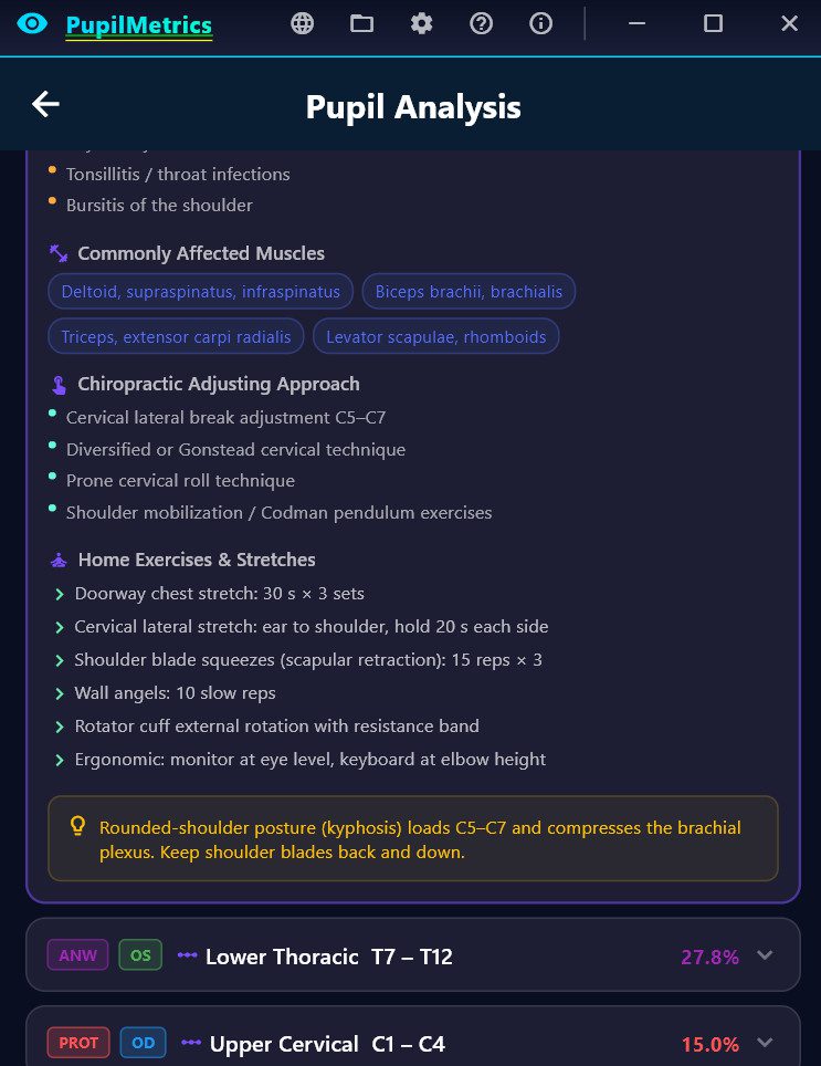
**What the panel shows**

Each spinal segment card displays:

| Section | Contents |

|———|———|

| **Segment label** | e.g. “Upper Cervical C1–C4” |

| **Vertebrae** | Individual vertebrae with common names (e.g. C1 Atlas, C2 Axis) |

| **Nerve roots** | Exiting nerve roots at that level |

| **Innervated structures** | Organs and tissues supplied by those nerves |

| **Subluxation indicators** | Classic symptoms associated with fixation at this level |

| **Affected muscles** | Muscles commonly involved in nerve compromise at this level |

| **Adjusting approach** | Standard chiropractic techniques applicable to the region |

| **Exercises** | Corrective exercises, stretches, and lifestyle recommendations |

| **Postural note** | Ergonomic or postural guidance specific to this spinal region |

**Sources**

Winsor (1921) sympathetic segmental disturbances study; Cleveland Chiropractic College nerve–organ chart; Palmer textbooks; Jensen and Angerer iridology references.

Updated on March 24, 2026

What are your Feelings

  • Happy
  • Normal
  • Sad

Share This Article :

  • Facebook
  • X
  • LinkedIn
  • Pinterest
5.4 Nutrition Recommendations Panel5.6 TCM Correlations Panel

The Clinical Neuro-Optic Research Initiative (CNRI) advances pupil-based neurodiagnostics by preserving historical insights, developing modern analytic tools, and researching links between ocular microstructures and systemic health. Our mission is to validate and expand neuro-optic biomarkers for breakthroughs in early detection, monitoring, and non-invasive assessment of autonomic and neurological function.

Twitter