Facebook Twitter VKontakte
Sign in
  • Home
  • Human Eye Project
    • The Pupil
    • Pupil Decentration-Multiformaties
    • Pupil Deformations
    • Pupil Color and Dimensions
    • Pupil Anisocoria
    • Pupil Miosis
    • Pupil Mydriasis
    • Pupil Reflexes
    • The Collarette
    • The Iris
  • PupilMetrics App
    • PupilMetrics Android
    • PupilMetrics Windows
    • PupilMetrics Mac OS
  • Documentation
  • Pricing
  • Learn More
    • CNRI Research
    • History
    • About
    • Eye Photo Tips & Tricks
    • IRB Status
    • Pupil Abstract Blog
    • Privacy Policy
    • Terms of Service
Sign in
Welcome!Log into your account
Forgot your password?
Password recovery
Recover your password
Search
Sign in
Welcome! Log into your account
Forgot your password? Get help
Password recovery
Recover your password
A password will be e-mailed to you.
Clinical Neuro-Optic Reseach Initiative
  • Home
  • Human Eye Project
    • The Pupil
    • Pupil Decentration-Multiformaties
    • Pupil Deformations
    • Pupil Color and Dimensions
    • Pupil Anisocoria
    • Pupil Miosis
    • Pupil Mydriasis
    • Pupil Reflexes
    • The Collarette
    • The Iris
  • PupilMetrics App
    • PupilMetrics Android
    • PupilMetrics Windows
    • PupilMetrics Mac OS
  • Documentation
  • Pricing
  • Learn More
    • CNRI Research
    • History
    • About
    • Eye Photo Tips & Tricks
    • IRB Status
    • Pupil Abstract Blog
    • Privacy Policy
    • Terms of Service

Getting Started

5
  • 1.1 System Requirements
  • 1.2 Installation
  • 1.3 Licensing
  • 1.4 First Launch
  • 1.5 Desktop Window & Keyboard Shortcuts

Capturing Eye Images

8
  • 2. Capturing Eye Images
  • 2.1 Camera Source Selection
  • 2.2 Quality-Gated Camera Mode – Android App
  • 2.3 Manual Camera Mode – Android
  • 2.4 USB / UVC Iriscope (Dino-Lite)
  • 2.5 PLR Video Mode – Android
  • 2.6 Import from Gallery
  • 2.7 Tips for a Good Capture

Reading the Analysis Results

10
  • 3. Reading the Analysis Results
  • 3.1 The Iris Zone Map
  • 3.2 PI Ratio (Pupil–Iris Ratio)
  • 3.3 Zone Findings — Flattenings (FLAT) and Protrusions (PROT)
  • 3.4 ANW Assessment (Collarette / Autonomic Nerve Wreath)
  • 3.5 Decentration (Pupil Position)
  • 3.6 Ellipseness (Pupil Shape)
  • 3.7 Anisocoria (Pupil Size Difference)
  • 3.8 Confidence Scores & Hybrid Fusion
  • 3.9 Scan History

Patient Management

4
  • 4. Patient Management & Exports
  • 4.1 PDF Report
  • 4.2 Plain-Text & JSON Export
  • 4.3 Sharing & Filing

Natural Medicine Therapy Panels

8
  • 5. Natural Medicine Therapy Panels
  • 5.1 Enabling the Therapy Modules
  • 5.2 How Zone Findings Drive the Therapy Panels
  • 5.3 Herbal Recommendations Panel
  • 5.4 Nutrition Recommendations Panel
  • 5.5 Chiropractic Correlations Panel
  • 5.6 TCM Correlations Panel
  • 5.7 Reading Therapy Panels Together

Constitutional Iridology

7
  • 6. Constitutional Iridology
  • 6.1 Background & Theoretical Basis
  • 6.2 The 34 Constitutional Types
  • 6.3 Selecting a Constitutional Type
  • 6.4 Constitutional Panel in Analysis Results
  • 6.5 Constitutional Section in the PDF Report
  • 6.6 Clinical Guidance & Limitations

Exporting PDF Reports

2
  • 7. Exporting PDF Reports
  • 7.1 Regenerating a PDF

Settings & Customization

5
  • 8. Settings & Customization
  • 8.1 Languages
  • 8.2 Zone Overlay & Observer Notes
  • 8.3 ML Comparison Panel
  • 8.4 About & Support

Clinical & Legal Disclaimers

2
  • 9. Clinical & Legal Disclaimers
  • 9.1 Data Privacy
View Categories
  • Home
  • PupilMetrics Documentation
  • Constitutional Iridology
  • 6.3 Selecting a Constitutional Type

6.3 Selecting a Constitutional Type

2 min read

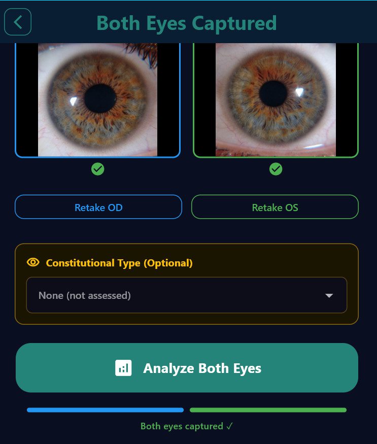
Constitutional type selection is performed on the **”Both Eyes Captured”** screen, which appears after both left and right eye photos have been taken. At this point the practitioner has both eye images visible side by side — the appropriate moment for constitutional assessment, since Deck’s system requires evaluation of both irides together.

**To select a type (Windows desktop only):**

1. Complete the right eye capture (Step 1 of 2).

2. Complete the left eye capture. The screen title changes to **”Both Eyes Captured”** when both images are present.

3. Scroll below the eye image pair. An amber-bordered panel labelled **”Constitutional Type (Optional)”** is visible.

4. Click the dropdown to open it. Types are organized by group with non-selectable group dividers.

5. Select the appropriate type. The type name is confirmed below the dropdown.

6. Leave the dropdown at **”None (not assessed)”** to omit the constitutional section entirely from the analysis and PDF.

7. Tap **Analyze Both Eyes** to proceed.

> **Session persistence:** The selected type is held in memory for the current session. If you navigate back to the Both-Eyes-Captured screen, the previously selected type is restored. The selection is cleared when a new scan session begins from the home screen.

**Assessment approach:** Constitutional typing requires experience and ideally evaluation of the iris under magnification (slit lamp or iriscope). The practitioner should consider:

– **Iris base color** — blue-grey → Lymphatic group; dark brown → Haematogenic; light brown/mixed → Biliary/Mixed

– **Fiber density and texture** — tight/silk-like vs. loose/wavy vs. coarsely woven with lacunae

– **Tophi and plaques** — presence, definition, and color of connective-tissue deposits

– **Pigmentation pattern** — central heterochromia, scurf rim, liver-zone pigments, dispersed spots

– **Special structures** — cramp rings, heart-zone defect markings, arcus senilis/lipaemic ring

When the constitutional group is clear but the exact subtype is uncertain, selecting the base type (e.g., *Pure Lymphatic*, *Classic Biliary*, *Haematogenic I*) is preferable to guessing a complex subtype.

Updated on March 24, 2026

What are your Feelings

  • Happy
  • Normal
  • Sad

Share This Article :

  • Facebook
  • X
  • LinkedIn
  • Pinterest
6.2 The 34 Constitutional Types6.4 Constitutional Panel in Analysis Results

The Clinical Neuro-Optic Research Initiative (CNRI) advances pupil-based neurodiagnostics by preserving historical insights, developing modern analytic tools, and researching links between ocular microstructures and systemic health. Our mission is to validate and expand neuro-optic biomarkers for breakthroughs in early detection, monitoring, and non-invasive assessment of autonomic and neurological function.

Twitter