Facebook Twitter VKontakte
Sign in
  • Home
  • Human Eye Project
    • The Pupil
    • Pupil Decentration-Multiformaties
    • Pupil Deformations
    • Pupil Color and Dimensions
    • Pupil Anisocoria
    • Pupil Miosis
    • Pupil Mydriasis
    • Pupil Reflexes
    • The Collarette
    • The Iris
  • PupilMetrics App
    • PupilMetrics Android
    • PupilMetrics Windows
    • PupilMetrics Mac OS
  • Documentation
  • Pricing
  • Learn More
    • CNRI Research
    • History
    • About
    • Eye Photo Tips & Tricks
    • IRB Status
    • Pupil Abstract Blog
    • Privacy Policy
    • Terms of Service
Sign in
Welcome!Log into your account
Forgot your password?
Password recovery
Recover your password
Search
Sign in
Welcome! Log into your account
Forgot your password? Get help
Password recovery
Recover your password
A password will be e-mailed to you.
Clinical Neuro-Optic Reseach Initiative
  • Home
  • Human Eye Project
    • The Pupil
    • Pupil Decentration-Multiformaties
    • Pupil Deformations
    • Pupil Color and Dimensions
    • Pupil Anisocoria
    • Pupil Miosis
    • Pupil Mydriasis
    • Pupil Reflexes
    • The Collarette
    • The Iris
  • PupilMetrics App
    • PupilMetrics Android
    • PupilMetrics Windows
    • PupilMetrics Mac OS
  • Documentation
  • Pricing
  • Learn More
    • CNRI Research
    • History
    • About
    • Eye Photo Tips & Tricks
    • IRB Status
    • Pupil Abstract Blog
    • Privacy Policy
    • Terms of Service

Getting Started

5
  • 1.1 System Requirements
  • 1.2 Installation
  • 1.3 Licensing
  • 1.4 First Launch
  • 1.5 Desktop Window & Keyboard Shortcuts

Capturing Eye Images

8
  • 2. Capturing Eye Images
  • 2.1 Camera Source Selection
  • 2.2 Quality-Gated Camera Mode – Android App
  • 2.3 Manual Camera Mode – Android
  • 2.4 USB / UVC Iriscope (Dino-Lite)
  • 2.5 PLR Video Mode – Android
  • 2.6 Import from Gallery
  • 2.7 Tips for a Good Capture

Reading the Analysis Results

10
  • 3. Reading the Analysis Results
  • 3.1 The Iris Zone Map
  • 3.2 PI Ratio (Pupil–Iris Ratio)
  • 3.3 Zone Findings — Flattenings (FLAT) and Protrusions (PROT)
  • 3.4 ANW Assessment (Collarette / Autonomic Nerve Wreath)
  • 3.5 Decentration (Pupil Position)
  • 3.6 Ellipseness (Pupil Shape)
  • 3.7 Anisocoria (Pupil Size Difference)
  • 3.8 Confidence Scores & Hybrid Fusion
  • 3.9 Scan History

Patient Management

4
  • 4. Patient Management & Exports
  • 4.1 PDF Report
  • 4.2 Plain-Text & JSON Export
  • 4.3 Sharing & Filing

Natural Medicine Therapy Panels

8
  • 5. Natural Medicine Therapy Panels
  • 5.1 Enabling the Therapy Modules
  • 5.2 How Zone Findings Drive the Therapy Panels
  • 5.3 Herbal Recommendations Panel
  • 5.4 Nutrition Recommendations Panel
  • 5.5 Chiropractic Correlations Panel
  • 5.6 TCM Correlations Panel
  • 5.7 Reading Therapy Panels Together

Constitutional Iridology

7
  • 6. Constitutional Iridology
  • 6.1 Background & Theoretical Basis
  • 6.2 The 34 Constitutional Types
  • 6.3 Selecting a Constitutional Type
  • 6.4 Constitutional Panel in Analysis Results
  • 6.5 Constitutional Section in the PDF Report
  • 6.6 Clinical Guidance & Limitations

Exporting PDF Reports

2
  • 7. Exporting PDF Reports
  • 7.1 Regenerating a PDF

Settings & Customization

5
  • 8. Settings & Customization
  • 8.1 Languages
  • 8.2 Zone Overlay & Observer Notes
  • 8.3 ML Comparison Panel
  • 8.4 About & Support

Clinical & Legal Disclaimers

2
  • 9. Clinical & Legal Disclaimers
  • 9.1 Data Privacy
View Categories
  • Home
  • PupilMetrics Documentation
  • Settings & Customization
  • 8. Settings & Customization

8. Settings & Customization

1 min read

Settings are stored via SharedPreferences and persist across app restarts. On Windows they survive app updates. There is no dedicated Settings screen — all settings are accessible from the **title-bar menu** (Windows) or the **main menu** on mobile.

Complete Settings Reference
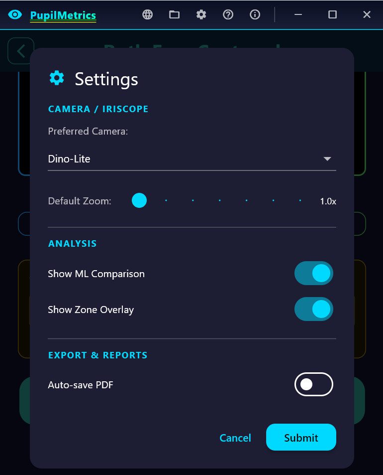
#### Camera & Capture

| Setting | Default | Options / Range | Effect |

|———|———|—————–|——–|

| **Preferred camera** | Dino-Lite | `dino_lite`, `usb_camera`, `auto_detect` | Pre-selects the camera source on the camera mode selector page |

| **Default zoom** | 1.0× | 1.0× – 4.0× | Starting zoom level applied when the standard camera opens |

The zoom slider maps a stored value of 0.0–1.0 to a display range of 1.0×–4.0× (formula: `display = 1.0 + stored × 3.0`). Setting zoom to 0 stored = 1.0× display (no zoom).
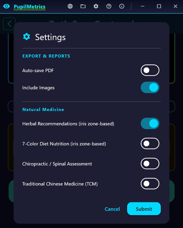
#### Report & PDF

| Setting | Default | Effect |

|———|———|——–|

| **Auto-save PDF** | Off | Save PDF automatically at end of every analysis |

| **Include images in PDF** | On | Embed OD/OS photos in the PDF; turn off to reduce file size |

#### Analysis Display

| Setting | Default | Effect |

|———|———|——–|

| **Show ML comparison** | On | Displays the ML model’s raw output values alongside the classical CV result on the results screen, for practitioner reference |

| **Show zone overlay** | On | Enables the interactive polar zone overlay on the iris photo on the results screen; tap zones to see finding details and add observer notes |

#### Practice Information

| Setting | Default | Effect |

|———|———|——–|

| **Practice / Clinic name** | *(empty)* | Text entered here (or on the patient info form) appears as a teal banner in every report header |

#### Natural Medicine Modules

| Setting | Default | Effect |

|———|———|——–|

| **Herbal mode** | Off | Enable herbal recommendation panel and PDF section |

| **Nutrition mode** | Off | Enable 7-color diet nutrition panel and PDF section |

| **Chiropractic mode** | Off | Enable chiropractic spinal correlation panel and PDF section |

| **TCM mode** | Off | Enable Traditional Chinese Medicine meridian panel and PDF section |

All four therapy toggles are independent. Enable only the modalities relevant to your practice.

Updated on March 24, 2026

What are your Feelings

  • Happy
  • Normal
  • Sad

Share This Article :

  • Facebook
  • X
  • LinkedIn
  • Pinterest
8.4 About & Support8.1 Languages

The Clinical Neuro-Optic Research Initiative (CNRI) advances pupil-based neurodiagnostics by preserving historical insights, developing modern analytic tools, and researching links between ocular microstructures and systemic health. Our mission is to validate and expand neuro-optic biomarkers for breakthroughs in early detection, monitoring, and non-invasive assessment of autonomic and neurological function.

Twitter